- The cervical spine is the most superior portion of the vertebral column, lying between the cranium and the thoracic vertebrae. It consists of seven vertebrae, two of which are given unique names:
- The first cervical vertebrae (C1) is known as the
- The second cervical vertebrae (C2) is known as the
- Upper cervical: C1, C2
- Lower cervical: C3 to C7
Features:
- C1 and C2are considered atypical vertebrae because they have some distinguishing features compared to the rest of the cervical spine. The top vertebra, called the atlas, is the only cervical vertebra without a vertebral body. Instead, it is shaped more like a ring.
- Triangular vertebral foramen.
- Transverse foramina– holes in the transverse processes. They give passage to the vertebral artery, vein and nerves.
- Bifid spinous process– this is where the spinous process splits into two distally.
Atlas(C1):
- Doesn’t Have body & spinous process
- Its ring-like, has anterior and a posterior arch and two lateral masses.
- Each lateral mass has superior articular facet & inferior articular facet.
- Superior articular facet articulate with occipital condoyle- atlanto-occipital joint.
- Inferior articular facet articulate with axis superior facet -atlanto-axis joint. Transverse process project laterally from lateral mass which is pierced by foramen transversorium.
- The vertebral body of C1 fuses onto the body of C2 during development to become the dens of C2.
- As a result, there is no intervertebral disc between Atlas and Axis.

Axis(C2):
- The second cervical vertebra (C2) of the spine is named the axis.
- The most distinctive characteristic of this bone is the strong odontoid process (“dens”) which rises perpendicularly from the upper surface of the body.

Joints:
Atlanto-occipital Joint:
The atlanto-occipital articulations function as bilaterally symmetrical synovial joints between C0 and C1. Articulation occurs at Occipital condyles and a superior articular facets of the atlas. The atlanto-occipital joint (synovial joint) allows the head to nod up and down on the vertebral column. Motion at the occiput-C1 segment is restricted primarily to flexion- extension due to bony structures, ligamentous constraints, and the absence of an intervertebral disc.

Atlantoaxial Joint:
JOINT between the atlas (C1) and the axis (C2); has a range of motion in the transverse plane for rotation. The DENS of C2 acts as a pivot point for the rotation of C1. The articulating surfaces of the two vertebrae form ZYGAPOPHYSEAL (FACET) JOINTS that allow flexion- extension, side bending, and rotational movements.

Movements:
- Approximately 50% of flexion- extension motion occurs at occiput-C1.
- Approximately 50% of rotation occurs at C1-C2.
- Lesser amounts of flexion- extension, rotation, and lateral bending occur segmentally between C2-C7.
Ligaments:
Anterior longitudinal ligament (ALL):
Attaches to and covers the anterior and lateral aspects of the vertebral bodies and intervertebral discs. From C2 to C1 it is known as the anterior atlantoaxial membrane, and from C1 to the anterior aspect of the foramen magnum as the anterior atlanto-occipital membrane.These are the only spinal ligaments that limit hyperextension of the vertebral column.

Posterior longitudinal ligament (PLL):
Is a much narrower ligament that runs along the posterior aspect of the vertebral bodies. From C2, the PLL strengthens and becomes known as the tectorial membrane. It then passes through the foramen magnum to attach to the floor of the cranial cavity. Functionally, it weakly resists hyperflexion of the vertebral column, and helps prevent posterior herniations of the nucleus pulposus.

Ligamentum flavum:
Runs between the lamina of adjacent vertebrae. As with the ALL, the ligamentum flava continues from C2 and C1 as the posterior atlanto-axial and atlanto-occipital membranes. These membranes limit excessive motion at the atlanto-occipital joints.

Ligaments unique to cervical spine:
Alar ligament: Runs from the posterior aspect of the dens of C2 to the lateral margins of the foramen magnum.

Apical ligament: Connects the tip or apex of the dens of C2 to the anterior aspect of the foramen magnum.

Transverse ligament: Arises from the tubercle on the atlas (C1) and holds the odontoid process firmly against the anterior arch. The transverse ligament is the primary stabilizer that prevents abnormal anterior translation of C1 on C2.

Muscles:
Anterior Muscles of Neck:
Rectus capitis anterior:
Origin: from the anterior surface of lateral mass of atlas.
Insertion: inserts to the inferior surface of basilar part of occipital bone.
Function: flex the head on the neck at the atlantooccipital joint and to stabilize the atlantooccipital joint.

Rectus capitis lateralis:
Origin: arises from the upper surface of the transverse process of the atlas.
Insertion: into the occipital bone.
Function: stabilize the atlanto-occipital joint during movement.

Longus Capitis:
Origin: originates from its inferior aspect, run from the transverse processes of the third, fourth, fifth and sixth cervical vertebrae.
Insertion: on the basilar part of occipital bone.
Muscles: Flex the head.

Rectus Capitis Posterior Major:
Originates from the spinous process of the C2 vertebrae and inserts into the occipital bone
Rectus Capitis Posterior Minor:
Originates from the posterior tubercle of atlas (C1) and insert on occipital bone.
Oblique Capitis Inferior:
Originating from the lateral surface of the spinous process of the axis insert onto the of the transverse process of the atlas.
Oblique Capitis Superior:
Orginates from Transverse process of atlas and insert on occipital bone.
Function: It extends and rotate head.
Cervical plexus:
Arises from the anterior rami of the spinal nerves associated with C1-C4 (some texts include the C5 nerve root). This plexus lies deep to the sternocleidomastoid (SCM) muscle and levator scapulae muscle. The branches of the cervical plexus can be categorized as either muscular or cutaneous.


About Authors
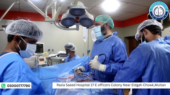
Dr. Muhammad Mahmood Ahmad is a Spinal as well as an Orthopedic Surgeon with over 14 years of experience currently practicing at Razia Saeed Hospital, Multan.