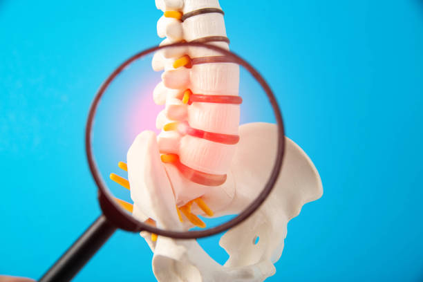
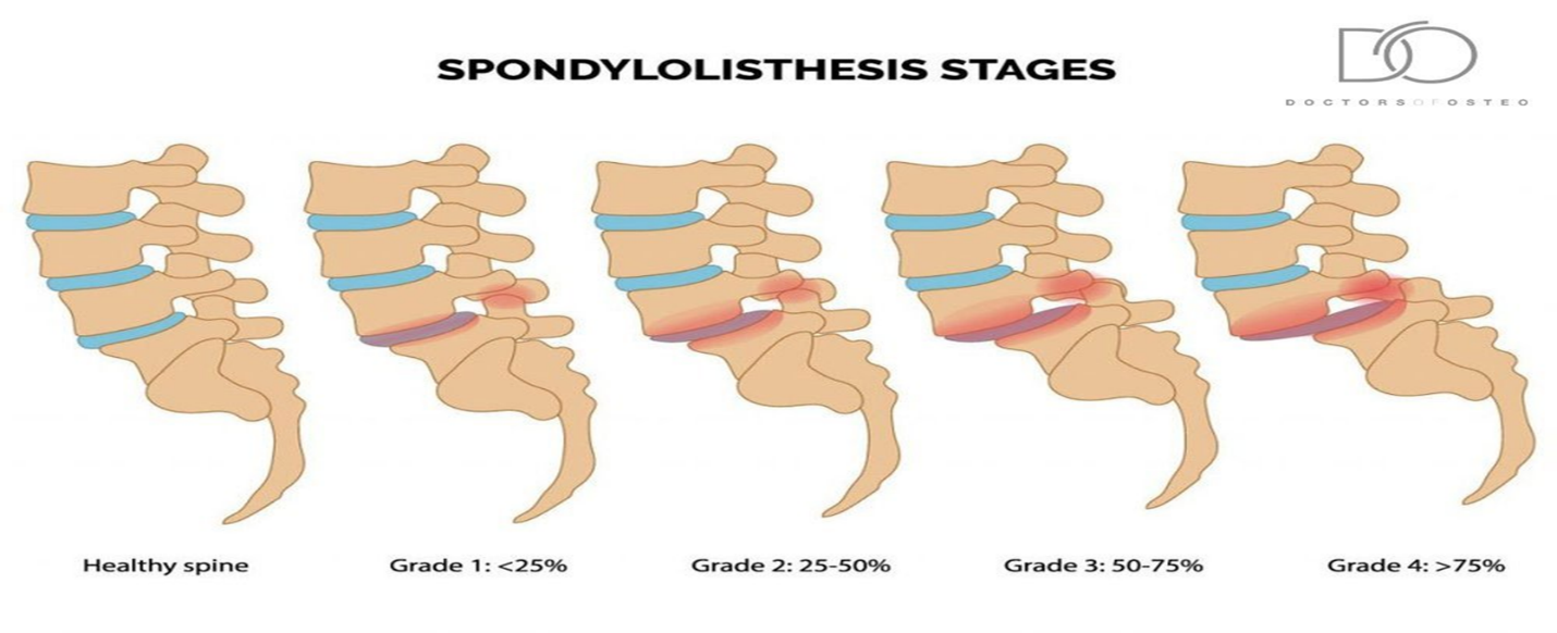
Spondylolisthesis is the slippage of one vertebral body with respect to the adjacent vertebral body causing mechanical or radicular symptoms or pain. It can be due to congenital, acquired, or idiopathic causes. Spondylolisthesis is graded based on the degree of slippage of one vertebral body on the adjacent vertebral body.
Stages:
Clinical Anatomy:
- Spondylolisthesis most commonly occurs in the lower lumbar spine but can also occur in the cervical spine and rarely, except for trauma, In the thoracic spine.
The pathology involves:
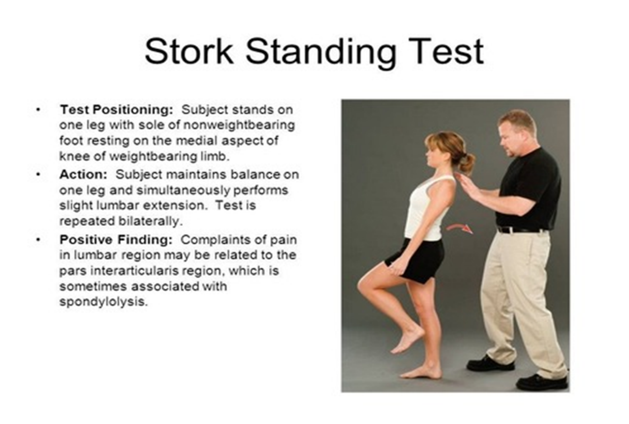
- A fractured pars interarticularis of the lumbar vertebrae, also called the isthmus.
- This affects the supporting structural integrity of the vertebrae, which could lead to slippage of the corpus of the vertebrae.
- In turn, leads to one of the most obvious manifestations of lumbar instability.
- Slippage can occur in 2 directions- most commonly in anterior translation, called anterolisthesis, or a backward translation, called retrolisthesis.
Etiology:
- Repetitive stress to the pars interarticularis.
- Traumatic accidental injuries.
- Microtrauma in sports.
- Pathological causes – Neoplasm, connective tissue disease, etc.
- Iatrogenic – After laminectomy.
- Adolescents and children also have more elastic intervertebral disks which cause increased stress to be placed on the pars interarticularis.
Wiltse Classification
Dysplastic Spondylolisthesis (Type I):
Dysplastic Spondylolisthesis is congenital and secondary to variation in the orientation of the facet joints to an abnormal alignment. In dysplastic spondylolisthesis, the facet joints are more sagittally oriented than the typical coronal orientation.
Isthmic Spondylolisthesis (Type II):
Isthmic spondylolisthesis results from defects in the pars interarticularis. The cause of isthmic spondylolisthesis is undetermined, but a possible etiology includes microtrauma in adolescence related to sports such as wrestling, football, and gymnastics, where repeated lumbar extension occurs. It has two types. Type IIA is caused by a stress fracture of the pars interarticularis (spondylolysis) that results in anterior slippage of the vertebrae. Type II B is caused by repetitive fractures and subsequent healing which results in lengthening of the pars interarticularis leading to anterior slippage of the vertebrae.
Degenerative Spondylolisthesis (Type III):
Degenerative spondylolisthesis (Type 3) occurs from degenerative changes in the spine without any defect in the pars interarticularis. It is usually related to combined facet joint and disc degeneration leading to instability and forward movement of one vertebral body relative to the adjacent vertebral body. Arthritis of facet joint which in turn causes weakness of ligamentum flavum leads to anterior slippage of vertebra.
Traumatic Spondylolisthesis (Type IV):
Traumatic Spondylolisthesis occurs after fractures of the pars interarticularis or the facet joint structure and is most common after trauma.
Pathologic Spondylolisthesis (Type V):
Pathologic Spondylolisthesis can be from systemic causes such as bone or connective tissue disorders or a focal process, including infection, neoplasm.
Iatrogenic Spondylolisthesis (Type VI):
Iatrogenic Spondylolisthesis is a potential sequela of spinal surgery. Frequently, these patients will have undergone prior laminectomy.
Clinical Presentation:
- Patients typically have low back pain which mimics radiculopathy for lumbar spondylolisthesis.
- Pain is exacerbated by extending at the affected segment, as this can cause mechanic pain from motion, leading to diminished ROM (spine).
- Pain decreases as the patient assumes flexed posture which reduces stress on the nerve being impinged.
- Pain may be exacerbated by direct palpation of the affected segment.
- Pain can also be radicular in nature as the exiting nerve roots become compressed due to the narrowing of nerve foramina as one vertebra slips on the adjacent vertebrae.
- Pain can sometimes improve in certain positions such as lying supine.
- Atrophy of the muscles, muscle weakness.
- Disturbances in coordination and balance, difficulty in walking.
Differential Diagnosis:
- Spondylolysis
- Metastatic disease
- Low back pain
- Osteoarthritis
- Neuroforaminal stenosis
- Spinal Stenosis
Diagnostic Procedures:
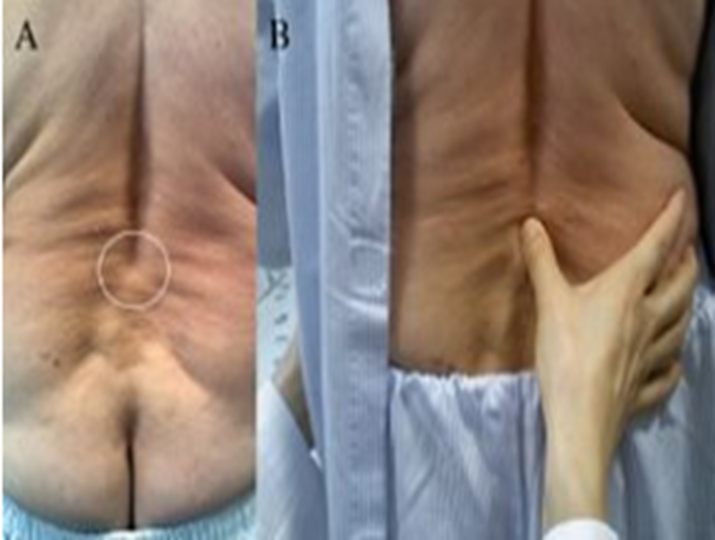
Step off Sign:
A noticeable Step off sign is palpated at the Lumbo sacral area due to slippage of the vertebrae.
Conservative Treatment:
- Initially resting and avoiding movements like lifting, bending, and sports.
- A brace or lumbosacral belt may be useful to decrease segmental spinal instability and pain.
- Physiotherapy focuses on relieving extension stresses from the lumbosacral junction (hamstring and hip flexor stretching), as well as working on core strengthening (deep abdominal muscles and lumbar multifidus strengthening.
Physical Therapy Protocol:
- Isometric and isotonic exercises beneficial for strengthening of the main muscles of the trunk, which stabilize the spine.
- Core stability exercises, useful in reducing pain and disability in chronic low back pain in patient with spondylolisthesis.
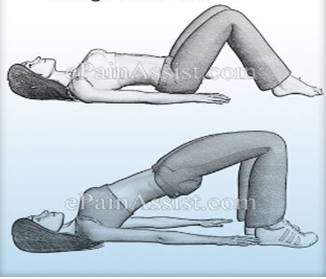
- Movements in closed-chain-kinetics like squats, lunges, push ups, hip bridge.
- Antilordotic movement patterns of the spine like dead bugs, sitting pelvic tilts, hamstring curl.
- Gait training.
- Stretching and strengthening exercises, objective of stretching and strengthening is to decrease the extension forces on the lumbar spine, due to agonist muscle tightness, antagonist weakness, or both, which may result in decreased lumbar lordosis.
- People with spondylolisthesis often have tight hamstrings, In order to improve the patient’s mobility stretching exercises of the hamstrings, hip flexors and lumbar paraspinal muscles are important.
- Balance training including – Sensomotoric training on devices, walking in all variations, coordinative skills.
- Endurance training of muscles, effective for chronic low back pain.
- Cardiovascular exercise – Athletes with spondylolysis and first-degree spondylolisthesis can take part in all sports activities.
Williams flexion exercises are a set of exercises that decrease lumbar extension and focuses on lumbar flexion. These include
- Pelvic Tilts
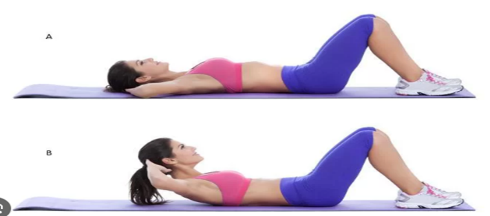
- Partial sit-ups
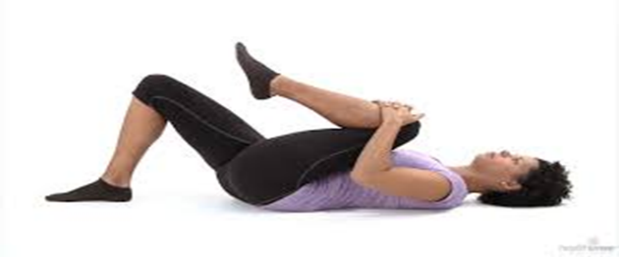
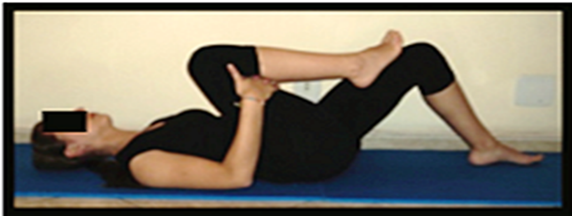
- Knee-to-chest
- Hamstring stretch
- Standing lunges
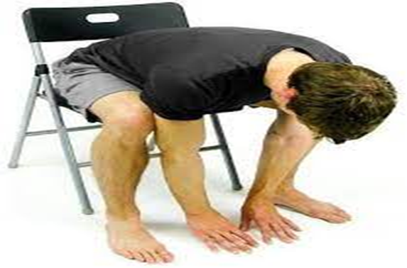
- Seated trunk flexion
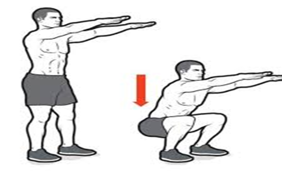
- Full squats
Pelvic Tilt Exercise
Partial Sit-ups
Knee to Chest Exercise
Hamstring stretch Exercise
Standing Lunges
Seated Trunk Flexion
Full Squats
Strengthening of the Deep Abdominal Muscles:
Alternating legs, with leg extension while exhaling, maintaining contraction of transverse abdominis, paravertebral and pelvic floor muscles.
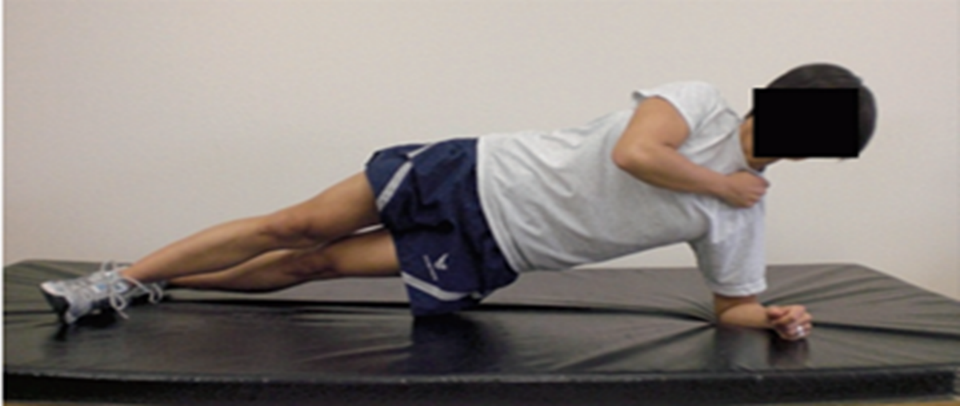
Horizontal side support exercise for core stability:
Stretching of the erector spine muscles:
About Authors
Dr. Muhammad Mahmood Ahmad is a Spinal as well as an Orthopedic Surgeon with over 14 years of experience currently practicing at Razia Saeed Hospital, Multan.
